Funded by the FWF doc.funds project Mohammad is doing his PhD at the Carinthia University of Applied Sciences in collaboration with the Medical University of Vienna as well as the Massachusetts General Hospital. Together, the teams investigated how modern AI methods can be combined with advanced MRI hardware to address a long-standing challenge in medical imaging.
The published paper can be accessed via the following link: https://www.nature.com/articles/s41598-026-56900-z
Keeping MRI Images Accurate – Even When We Move
Anyone who has undergone an MRI scan has probably heard the instruction: “Please stay as still as possible.” Unfortunately, even small head movements can affect image quality and reduce the accuracy of advanced MRI examinations.
MRI scanners rely on an extremely uniform main magnetic field. In reality, however, the main magnetic field is never perfectly homogeneous. Differences between bone, air cavities, and brain tissue create local distortions in the magnetic field. To compensate for these distortions, MRI systems use a process called shimming. In simple terms, shimming adjusts additional magnetic fields to make the overall magnetic field uniform. The challenge with that is that shimming is usually performed only once at the beginning of a scan. When a person moves, the magnetic field changes, and the previously optimized correction is no longer accurate. As a result, image distortions, signal loss, and reduced spectral quality can occur.
For my PhD project I am addressing this problem by combining advanced hardware with artificial intelligence, as explained further below.
Combining Advanced Hardware and Artificial Intelligence
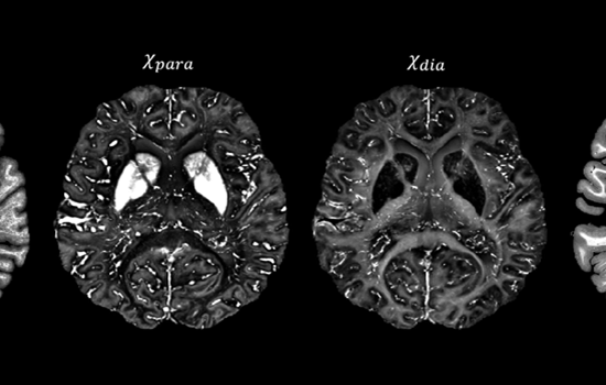
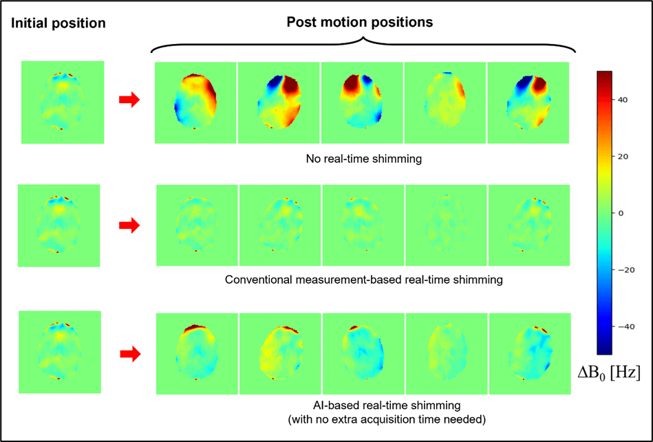
The project investigated a combination of conventional MRI shim coils and an advanced multi-coil technology known as AC/DC matrix coils. While these coils can significantly improve magnetic field homogeneity, their benefits are reduced when patients move (Figure 1). Our simulations showed that maintaining these improvements requires continuous updates of the magnetic field correction.
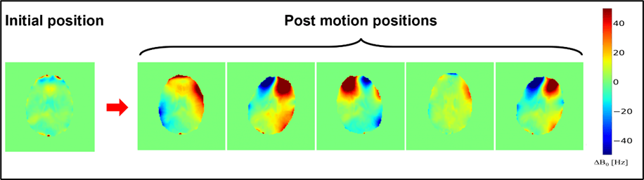
This is where AI becomes valuable: it can rapidly predict the required corrections and support real-time shimming without interrupting the main MRI scan. Now, instead of repeatedly measuring the magnetic field during the scan, a deep-learning model learns the relationship between head position and magnetic field distortions. The AI can then estimate these changes almost instantly and provide information needed for real-time correction (Figure 2).
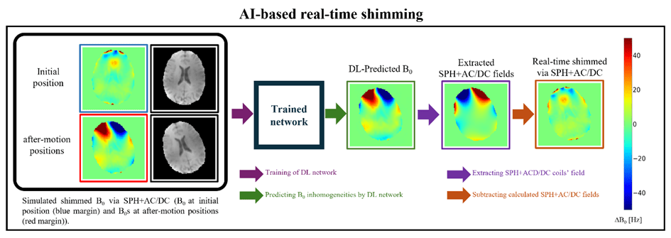
Our results showed that the performance was comparable to conventional measurement-based approaches (Figure 3), and that AI-based real-time shimming was able to maintain magnetic field homogeneity even when substantial head motion occurred. Although further validation on physical hardware is still required, our results demonstrate the potential of AI-assisted MRI systems that can automatically adapt to patient motion.
Yours,
MedTech @ FH Kärnten Team